Showing posts with label Alice's Treatment Room. Show all posts

Sunday, September 27, 2015

What Does Osteopathy Treat?

Osteopathy is a form of drug-free non-invasive manual medicine that focuses on total body health by treating and strengthening the musculoskeletal framework, which includes the joints, muscles and spine. Its aim is to positively affect the body's nervous, circulatory and lymphatic systems. 

This therapy is a unique holistic (whole body) approach to health care. Osteopaths do not simply concentrate on treating the problem area, but use manual techniques to balance all the systems of the body, to provide overall good health and wellbeing. 


Dr. Andrew Taylor Still established the practice of Osteopathy in the late 1800s in the United States of America, with the aim of using manual 'hands on' techniques to improve circulation and correct altered biomechanics, without the use of drugs. 

What does osteopathy treat?

Osteopaths treat more than you think........
Many patients present with complaints of aches in the head, back, neck, and heel/ foot pain; sciatica; shin splints; tennis elbow and repetitive strain injury. Other patients suffer from asthma; arthritis; digestive problems; carpal tunnel syndrome; whiplash and postural problems.
Osteopaths also deal regularly with patients who have been injured in the workplace, at home or while playing sport. 

What are the qualities of Osteopathy

The philosophy of Osteopathy is what sets it apart from other medical disciplines. The key principles are based on all parts of the body functioning together in an integrated manner. If one part of the body is restricted, then the rest of the body must adapt and compensate for this, eventually leading to inflammation, pain, stiffness and other health conditions. When the body is free of restrictions in movement, Osteopathic treatment assists the body with pain minimisation, reduced stress and greater mobility providing the body with the opportunity to heal itself. 

Osteopaths use a broad range of gentle hands-on techniques including soft tissue stretching, deep tactile pressure, and mobilisation or manipulation of joints.
In some cases, Osteopaths can complement the advice given by GPs. For example, people who suffer from arthritis are often prescribed medication by their GP. In addition to that, Osteopaths can ease the pain caused by joint and muscle stiffness, by improving joint mobility and the flow of blood to the joints, and show arthritis sufferers how to prevent causing injury to themselves. 

Osteopathy is a five-year university course, which includes a degree and masters qualification. Senior Osteopathy students complete clinical training under the supervision of registered Osteopaths at student teaching clinics. 

Osteopaths believe in working as part of a health system of health providers and often refer back to the G.P. or another allied health professional where appropriate. 

Benefits of Osteopathy

Osteopathic treatment in itself is not 'preventative'. Osteopaths respect the body's natural ability as a self-regulating mechanism and only intervene when pain or discomfort is present. The benefits of osteopathy are the general improvement in mobility and structural stability of the body. In turn, other systems of the body such as the circulatory, nervous and lymphatic systems function more effectively and for a number of general conditions, minimal treatment is required. 


 Ref:by Christian Nordqvist 


Wednesday, July 15, 2015

Myasthenia gravis(MG)

Myasthenia gravis is a rare long-term condition that causes certain muscles to become weak.

It mainly affects muscles that are controlled voluntarily  often those controlling eye and eyelid movement, facial expression, chewing, swallowing and speaking.
Sometimes, the muscles that control breathing, neck and limb movements are also affected. 
The muscle weakness associated with myasthenia gravis is usually worse during, or just after, physical activity and improves with rest. The symptoms are often described as being at their worst when a person is tired, for example, at the end of the day.

What causes myasthenia gravis?
Myasthenia gravis is an autoimmune condition that affects the nerves and muscles. Autoimmune conditions are caused by the body's immune system mistakenly attacking healthy tissue.In myasthenia gravis, the immune system produces antibodies (proteins) that block or damage muscle receptor cells.
This prevents messages being passed from the nerve endings to the muscles, which results in the muscles not contracting (tightening) and becoming weak.
It's not fully understood why some people's immune systems produce specific antibodies that block the nerve's signal to the muscle.

Diagnosing myasthenia gravis

The process of diagnosing myasthenia gravis can take a long time because muscle weakness is a common symptom of many different conditions.
Your GP will look at your medical history and symptoms. They may suspect myasthenia gravis if your eye movements are impaired or if you have muscle weakness but you're still able to feel things. Around half of people with myasthenia gravis initially develop symptoms of double vision or eyelid droop, with more than 90% of people developing these symptoms at some point during the illness.
You may be referred to a neurologist (specialist in nervous system disorders), who will carry out some tests to help confirm the diagnosis.


Treating myasthenia gravis

Although there's no cure for myasthenia gravis, treatments are available to help control the symptoms and improve muscle weakness. 
Medication can be used to improve communication between the nerves and muscles, and to increase muscle strength.
Immunosuppressants are a type of medication used to improve muscle strength by controlling the production of abnormal antibodies.
In some cases of myasthenia gravis, surgery may be recommended to remove the thymus gland (a thymectomy). The thymus gland is found underneath the breastbone and is part of the immune system. It's often abnormal in people with myasthenia gravis.


Some people may experience a temporary or permanent period of remission (where there are no longer symptoms) and treatment can be stopped.
Permanent remissions occur in about a third of all people who have a thymectomy. Removing a thymus gland tumour (thymoma) usually has little or no effect on the underlying myasthenia gravis.

Who is affected by myasthenia gravis?

Myasthenia gravis is a rare condition, affecting about 15 in every 100,000 people in the North of America.
It can develop at any age, but most commonly affects women under 40 years of age and men over 60. Cases of myasthenia gravis are increasing, particularly in people over 50, but the reasons for this increase aren't fully understood.

Sunday, March 15, 2015

Lung Collapse


A collapse of the lung, also called pneumothorax, occurs when air that normally circulates within the lung leaks into the space between the lung and the chest wall, pushing the lung down or causing it to collapse. This causes the affected section of the lung to shut down. In most cases, only a portion of the lung collapses, leaving the rest of the lung intact. 

A severe lung collapse can cause low blood oxygen levels, respiratory failure, cardiac arrest, and shock. This is a medical emergency and can be fatal if not treated immediately.

What Causes Lung Collapse?

A lung collapse can occur as the result of trauma to the chest, damaged lung tissue, or a malfunction within the lung itself.

Trauma

Pneumothorax is usually caused by trauma to the chest. Traumatic events can include:
  • a rib fracture
  • a gun shot wound
  • a knife wound
  • a hard blow to the chest
  • CPR (cardiopulmonary resuscitation)
  • a lung biopsy (using a needle through the chest)
  • endoscopy procedures

Damaged Lung Tissue

Damaged lung tissue can also cause lung collapse. Damaged tissue isn’t as strong as healthy tissue, so it collapses more easily. This damage can be a result of inflammatory or traumatic lung disease, but it can also be caused by: and
  • tuberculosis
  • pneumonia
  • lung cancer
  • sarcoidosis (a chronic, nodular inflammatory disease)
  • pulmonary fibrosis (the formation of excess connective tissue in the lungs)
  • cystic fibrosis (hereditary condition with buildup of thick mucus in lungs and other organs)

Air Blebs

Air blebs are small, air-filled blisters that form on the outer lining of your lungs. They are not a sign of any disease or other lung condition, and experts aren’t sure why they form in certain people. Sometimes, these air blisters rupture spontaneously or due to changes in air pressure and cause pneumothorax. 

When to See a Doctor About Your Symptoms

When pneumothorax first occurs, you may feel a sharp pain in your chest that gets worse if you cough. You may also have difficulty breathing or shortness of breath. These symptoms can cause mild discomfort in some patients and are often confused with the symptoms of a chest cold or bronchitis. 

If the collapse affects a larger portion of your lung, you may experience other symptoms in conjunction with chest pain and shortness of breath. 

These symptoms include:
  • tiring easily
  • a rapid heart beat
  • a tight feeling in the chest
  • bluish skin tone
  • low blood pressure
  • flared nostrils when breathing
All symptoms of pneumothorax should be treated as a medical emergency. Even if the symptoms are mild, seek urgent care as soon as possible.
Part 4 of 6: Diagnosis

Diagnosing Pneumothorax

The doctor will listen to your lungs with a stethoscope while asking you to breathe deeply and exhale. If your lung has collapsed, the doctor will have trouble hearing breathing sounds from the affected lung. If the doctor has trouble hearing your breathing sounds, he or she may order imaging tests to get a better look at your lungs. 

Doctors can easily identify a collapsed lung using a chest X-ray. Your doctor may also order an imaging test called a CT (computerized tomography) scan to view the affected lung in more detail.
Part 5 of 6: Treatment

Treating Lung Collapse

Treatments for a collapsed lung are designed to restore lung function by removing external pressure on the lung.

When Treatment is Unnecessary

If the lung collapse is mild or affects only a small portion of the lung, your doctor may choose to monitor it carefully rather than treat it. Throughout the course of observation you may have several chest X-rays. You may also be ordered to rest more to speed up the healing process. If your doctor feels it is necessary, he or she may give you supplemental oxygen to help your lungs recover.

Suctioning Off Excess Air

For collapses that affect a larger area of the lung, you will need treatment to remove the excess air from your chest cavity. The air may be removed using a chest tube or needle. A needle with a syringe is inserted into your chest near the area of collapse and in the area of your rib cage. Your doctor then pulls up the syringe on the needle to suction out the air. 

A chest tube is used in the same way, though it is usually connected to a water seal container or a machine that suctions the air continuously. If the area of collapsed lung is large, the machine may need to suction the area for several days.

Surgery

If an air leak from the lung is the underlying cause of the collapse, surgery may be necessary to repair the leak. Your doctors will make two small incisions in your chest and insert a fiber optic camera into one of them to view the lung. A surgical tool will be inserted into the other incision and used to close the leak.
If an air bleb is to blame, the surgeon will sew it closed. If no bleb is visible, he or she will instill a talc-like substance into the pleural space to cause irritation. The irritated will then seal itself together to close off the leak. 


 Written by April Kahn

Friday, February 20, 2015

Chinese Cupping Therapy

Cupping is one of the oldest methods of traditional Chinese medicine. The earliest recorded use of cupping dates to the early fourth century, when the noted herbalist Ge Hong wrote about a form of cupping in A Handbook of Prescriptions. Later books written during the Tang and Qing dynasties described cupping in great detail; one textbook included an entire chapter on “fire jar qi,” a type of cupping that could alleviate headaches, dizziness and abdominal pain.

Originally, practitioners would use hollowed-out animal horns for cups, and place them over particular points or meridians. Today, most acupuncturists use cups made of thick glass or plastic, although bamboo, iron and pottery cups are still used in other countries. Glass cups are the preferred method of delivery, because they do not break as easily as pottery or deteriorate like bamboo, and they allow the acupuncturist to see the skin and evaluate the effects of treatment.


How does cupping work? What does it treat?
In a typical cupping session, glass cups are warmed using a cotton ball or other flammable substance, which is soaked in alcohol, let, then placed inside the cup. Burning a substance inside the cup removes all the oxygen, which creates a vacuum.

As the substance burns, the cup is turned upside-down so that the practitioner can place the cup over a specific area. The vacuum created by the lack of oxygen anchors the cup to the skin and pulls it upward on the inside of the glass as the air inside the jar cools. Drawing up the skin is believed to open up the skin’s pores, which helps to stimulate the flow of blood, balances and realigns the flow of qi, breaks up obstructions, and creates an avenue for toxins to be drawn out of the body.

Depending on the condition being treated, the cups will be left in place from 5 to 10 minutes. Several cups may be placed on a patient’s body at the same time. Some practitioners will also apply small amounts of medicated oils or herbal oils to the skin just before the cupping procedure, which lets them move the cups up and down particular acupoints or meridians after they have been applied.


In China, cupping is used primarily to treat respiratory conditions such as bronchitis, asthma, and congestion; arthritis; gastrointestinal disorders; and certain types of pain. Some practitioners also use cupping to treat depression and reduce swelling. Fleshy sites on the body, such as the back and stomach (and, to a lesser extent, the arms and legs), are the preferred sites for treatment.

How many types of cupping are there?
In addition to the traditional form of cupping described above, which is known as “dry” cupping, some practitioners also use what is called “wet” or “air” cupping.
In “air” cupping, instead of using a flame to heat the cup, the cup is applied to the skin, and a suction pump is attached to the rounded end of the jar. The pump is then used to create the vacuum. In “wet” cupping, the skin is punctured before treatment. 

When the cup is applied and the skin is drawn up, a small amount of blood may flow from the puncture site, which are believed to help remove harmful substances and toxins from the body.

Is cupping safe? Does it hurt?
While cupping is considered relatively safe (especially air cupping, which does not include the risk of fire and heat), it can cause some swelling and bruising on the skin. As the skin under a cup is drawn up, the blood vessels at the surface of the skin expand. This may result in small, circular bruises on the areas where the cups were applied. These bruises are usually painless, however, and disappear within a few days of treatment.


In addition, there are several instances where cupping should not be performed. Patients with inflamed skin; cases of high fever or convulsions; and patients who bleed easily, are not suitable candidates for cupping. Pregnant women should not have cupping on their stomach or lower back. If the cups are being moved, they should not cross bony areas, such as the ridges of the spine or the shoulder blades.

Back Muscles spasm

Back Muscles spasm

Causes
Normal voluntary muscle contraction begins when electrical signals are sent from the brain through the spinal cord along nerve cells called motor neurons.

These include both the upper motor neurons within the brain and the lower motor neurons within the spinal cord and leading out to the muscle. At the muscle, chemicals released by the motor neuron stimulate the internal release of calcium ions from stores within the muscle cell. 

These calcium ions then interact with muscle proteins within the cell, causing the proteins (actin and myosin) to slide past one another. This motion pulls their fixed ends closer, thereby shortening the cell and, ultimately, the muscle itself. Recapture of calcium and unlinking of actin and myosin allows the muscle fiber to relax. 

Abnormal contraction may be caused by abnormal activity at any stage in this process. Certain mechanisms within the brain and the rest of the central nervous system help regulate contraction. Interruption of these mechanisms can cause spasm. Motor neurons that are overly sensitive may fire below their normal thresholds. The muscle membrane itself may be over sensitive, causing contraction without stimulation. Calcium ions may not be recaptured quickly enough, causing prolonged contraction. 

Interuption of brain mechanisms and overly sensitive motor neurons may result from damage to the nerve pathways. Possible causes include stroke, multiple sclerosis, cerebral palsy, neurodegenerative diseases, trauma, spinal cord injury, and nervous system poisons such as strychnine, tetanus, and certain insecticides. Nerve damage may lead to a prolonged or permanent muscle shortening called contracture. 


Changes in muscle responsiveness may be due to or associated with:

Prolonged exercise:
Curiously, relaxation of a muscle actually requires energy to be expended. The energy is used to recapture calcium and to unlink actin and myosin. 
Normally, sensations of pain and fatigue signal that it is time to rest. Ignoring or overriding those warning signals can lead to such severe energy depletion that the muscle cannot be relaxed, causing a cramp. The familiar advice about not swimming after a heavy meal, when blood flow is directed away from the muscles, is intended to avoid this type of cramp.
Dehydration and salt depletion:
This may be brought on by protracted vomiting or diarrhea, or by copious sweating during prolonged exercise, especially in high temperatures. Loss of fluids and salts—especially sodium, potassium, magnesium, and calcium—can disrupt ion balances in both muscle and nerves. This can prevent them from responding and recovering normally, and can lead to cramp.
Metabolic disorders that affect the energy supply in muscle. 
These are inherited diseases in which particular muscle enzymes are deficient. They include deficiencies of myophosphorylase (McArdle's disease), phosphorylase b kinase, phosphofructokinase, phosphoglycerate kinase, and lactate dehydrogenase.
Myotonia:
This causes stiffness due to delayed relaxation of the muscle, but does not cause the spontaneous contraction usually associated with cramps. However, many patients with myotonia do experience cramping from exercise. Symptoms of myotonia are often worse in the cold. 
Myotonias include myotonic dystrophy, myotonia congenita, paramyotonia congenita, and neuromyotonia.


Risk Factors

Anyone is at risk for muscle spasms. 


Symptoms

The pain of a muscle cramp is intense, localized, and often debilitating Coming on quickly, it may last for minutes and fade gradually. Contractures develop more slowly, over days or weeks, and may be permanent if untreated. Fasciculations may occur at rest or after muscle contraction, and may last several minutes. 


Diagnosis
Abnormal contractions are diagnosed through a careful medical history, physical and neurological examination, and electromyography of the affected muscles. Electromyography records electrical activity in the muscle during rest and movement. 


Treatment

Most cases of simple cramps require no treatment other than patience and stretching. Gently and gradually stretching and massaging the affected muscle may ease the pain and hasten recovery.
More prolonged or regular cramps may be treated with drugs such as carbamazepine, phenytoin, or quinine. Fluid and salt replacement, either orally or intravenously, is used to treat dehydration. Treatment of underlying metabolic or neurologic disease, where possible, may help relieve symptoms.

Alternative Treatment

Cramps may be treated or prevented with Gingko (Ginkgo biloba) or Japanese quince (Chaenomeles speciosa). Supplements of vitamin E, niacin, calcium, and magnesium may also help. Taken at bedtime, they may help to reduce the likelihood of night cramps. 

Prevention
The likelihood of developing cramps may be reduced by eating a healthy diet with appropriate levels of minerals, and getting regular exercise to build up energy reserves in muscle. Avoiding exercising in extreme heat helps prevent heat cramps. Heat cramps can also be avoided by taking salt tablets and water before prolonged exercise in extreme heat. 

Taking a warm bath before bedtime may increase circulation to the legs and reduce the incidence of nighttime leg cramps. 

Tuesday, July 29, 2014

Achilles tendinopathy

Your Achilles tendon is an important part of your leg. It is found just behind and above your heel. It joins your heel bone (calcaneum) to your calf muscles. The function of your Achilles tendon is to help in bending your foot downwards at the ankle. (This movement is called plantar flexion by doctors.)

Achilles tendinopathy is a condition that causes pain, swelling, stiffness and weakness of the Achilles tendon. It is thought to be caused by repeated tiny injuries (known as microtrauma) to the Achilles tendon. After each injury, the tendon does not heal completely, as should normally happen. This means that over time, damage to the Achilles tendon builds up and Achilles tendinopathy can develop.

There are a number of things that may lead to these repeated tiny injuries to the Achilles tendon. 
For example:
  • Overuse of the Achilles tendon. This can be a problem for people who run regularly. (Achilles tendinopathy can also be a problem for dancers and for people who play a lot of tennis or other sports that involve jumping.)
  • Training or exercising wearing inappropriate footwear.
  • Having poor training or exercising techniques - for example, a poor running technique.
  • Making a change to your training programme - for example, increasing the intensity of your training and how often you train.
  • Training or exercising on hard or sloped surfaces.
  • Having a high-arched foot.
  • Having poor flexibility - for example, having tight or underdeveloped thigh (hamstring) muscles.


Achilles tendinopathy is also more common in people who have certain types of arthritis, such as ankylosing spondylitis or psoriatic arthritis. It is also thought that your genetic 'makeup' (the material inherited from your parents which controls various aspects of your body) may play a part for some people who develop Achilles tendinopathy.

People who are taking medicines from a group called fluoroquinolones (eg, the antibiotics ciprofloxacin and ofloxacin) for long periods also have an increased risk of developing Achilles tendinopathy.

Achilles tendinopathy used to be known as Achilles tendonitis. In general, 'itis' usually refers to inflammation, so tendonitis would mean inflammation of a tendon. However, Achilles tendinopathy is now thought to be a better term to use because it is thought that there is little or no inflammation that causes the problem.

If the Achilles tendon is torn, this is called an Achilles tendon rupture. There is a separate leaflet called Achilles Tendon Rupture that discusses this in more detail. The rest of this leaflet is just about Achilles tendinopathy.


What are the symptoms of Achilles tendinopathy? The main symptoms include pain and stiffness around the affected Achilles tendon. Pain and stiffness tend to develop gradually and are usually worse when you first wake up in the morning. (Severe pain that comes on suddenly and difficulty walking can be symptoms of Achilles tendon rupture. See a doctor urgently if you develop these symptoms.)
Some people have pain during exercise but, in general, pain is worse after exercise. Runners may notice pain at the beginning of their run, which then tends to ease and become more bearable, followed by an increase in pain when they have stopped running. Pain due to Achilles tendinopathy may actually prevent you from being able to carry out your usual everyday activities such as walking to the shops, etc. You may notice that you have pain when you touch the area around your Achilles tendon. There may also be some swelling around this area.

Diagnose:
usually diagnose Achilles tendinopathy because of your typical symptoms and from examining your Achilles tendon. They may feel for swelling or tenderness of the tendon. They may also ask you to do some exercises to put some stress on your Achilles tendon. 

For example, they may ask you to stand on the affected leg and raise your heel off the ground. For most people with Achilles tendinopathy this movement brings on (reproduces) their pain. 

If this does not reproduce pain, your doctor may ask you to hop on that foot, either on the spot or in a forwards direction. Your doctor may also do some other tests to make sure that there are no signs that you have ruptured your Achilles tendon. For example, squeezing your calf muscles and looking at how your foot moves.

X-rays or other tests are not usually needed to diagnose Achilles tendinopathy. However, an ultrasound scan or an MRI scan may sometimes be suggested by a specialist if the diagnosis is not clear.
What is the initial treatment for Achilles tendinopathy?
There are a number of treatments that may help. The treatments below are usually suggested first. 

They are all considered as conservative treatments. This means treatments that do not involve surgery.

Rest:

Rest and time off from sporting activities are important if you have Achilles tendinopathy. At first, you should stop any high-impact activities or sports (such as running). As pain improves, you can restart exercise as your pain allows. 

It is thought that complete rest, if it is prolonged, can actually be worse for the injury. Talk to your doctor about when you should start exercising again.

Ice packs:
Ice treatment may be useful for pain control and may help to reduce swelling in the early stages of Achilles tendinopathy. An ice pack should be applied for 10-30 minutes. Less than 10 minutes has little effect. More than 30 minutes may damage the skin. Make an ice pack by wrapping ice cubes in a plastic bag or towel. (Do not put ice directly next to skin, as it may cause ice burn.) A bag of frozen peas is an alternative. 

Gently press the ice pack on to the injured part. The cold from the ice is thought to reduce blood flow to the damaged tendon. This may limit pain and inflammation. Do not leave ice on while asleep.

Achilles tendon exercises

Some special exercises to help to stretch and strengthen your Achilles tendon can be helpful. You should aim to do these every day. Such exercises may help with pain control and stiffness.

A physiotherapist may be able to help you with these exercises as needed. They may also use other treatments such as ultrasound and massage to help relieve symptoms and promote healing of your Achilles tendon.

The following exercises can be used to help treat Achilles tendinopathy:

Monday, June 23, 2014

Morton’s Neuroma

 
If you sometimes feel that you are "walking on a marble," and you have persistent pain in the ball of your foot, you may have a condition called Morton's neuroma. A neuroma is a benign tumor of a nerve. Morton's neuroma is not actually a tumor, but a thickening of the tissue that surrounds the digital nerve leading to the toes.

Definition 
Morton's neuroma occurs as the nerve passes under the ligament connecting the toe bones (metatarsals) in the forefoot.
Morton's neuroma most frequently develops between the third and fourth toes, usually in response to irritation, trauma or excessive pressure. 


The incidence of Morton's neuroma is 8 to 10 times greater in women than in men. 

Symptoms
  • Normally, there are no outward signs, such as a lump, because this is not really a tumor.
  • Burning pain in the ball of the foot that may radiate into the toes. The pain generally intensifies with activity or wearing shoes. Night pain is rare.
  • There may also be numbness in the toes, or an unpleasant feeling in the toes.
Runners may feel pain as they push off from the starting block. High-heeled shoes, which put the foot in a similar position to the push-off, can also aggravate the condition. Tight, narrow shoes also aggravate this condition by compressing the toe bones and pinching the nerve. 

Risk factors 

Factors that appear to contribute to Morton's neuroma include:

High heels:
Wearing high-heeled shoes or shoes that are tight or ill fitting can place extra pressure on your toes and the ball of your foot. 
Certain sports:
Participating in high-impact athletic activities such as jogging or running may subject your feet to repetitive trauma. Sports that feature tight shoes, such as snow skiing or rock climbing, can put pressure on your toes. 
Foot deformities:
People who have bunions, hammertoes, high arches or flatfeet are at higher risk of developing Morton's neuroma.

Diagnosis 
During the examination, your physician will feel for a palpable mass or a "click" between the bones. He or she will put pressure on the spaces between the toe bones to try to replicate the pain and look for calluses or evidence of stress fractures in the bones that might be the cause of the pain. 


Range of motion tests will rule out arthritis or joint inflammations. 

X-rays may be required to rule out a stress fracture or arthritis of the joints that join the toes to the foot.


What is the standard examination in a doctor’s office to confirm a Morton’s neuroma? - See more at: http://teamdoctorsblog.com/2012/12/mortons-neuroma-self-help-tips-treatment-and-prevention-from-the-barefoot-running-doctor-at-team-doctors/#sthash.W2UWjOQ3.dpuf
What is the standard examination in a doctor’s office to confirm a Morton’s neuroma?

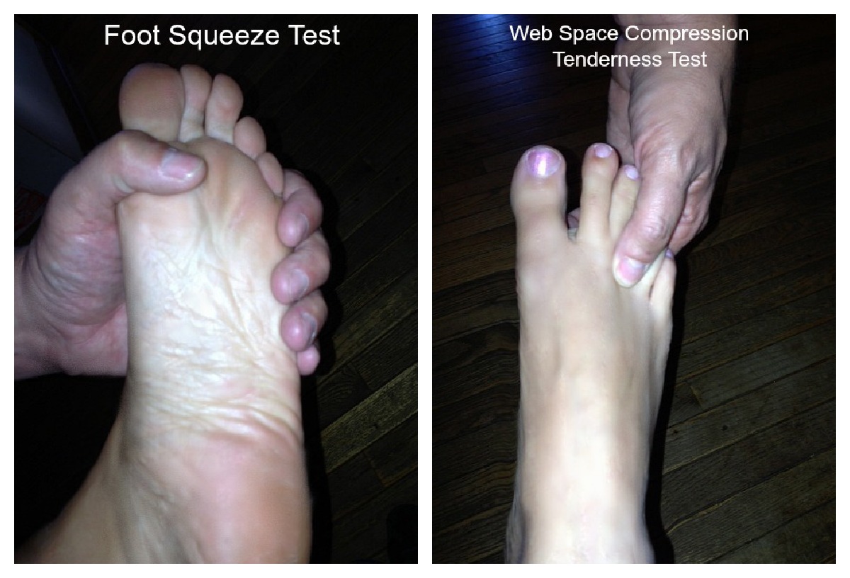
Morton’s Neuroma Tests
Web Space Compression Tenderness Test – Doctors push their thumb between the 3rd and 4th toes (metatarsals). If there is pain there they suspect a Morton’s neuroma.
Foot Squeeze Test – Doctors squeeze the foot from the sides and if it hurts between the bones, they suspect a Morton’s neuroma. (8)
Gauthers Test - involves squeezing the metatarsals together and moving them up and down for 30 seconds, which leads to pain.
- See more at: http://teamdoctorsblog.com/2012/12/mortons-neuroma-self-help-tips-treatment-and-prevention-from-the-barefoot-running-doctor-at-team-doctors/#sthash.W2UWjOQ3.dpuf
Treatment 
Initial therapies are nonsurgical and relatively simple. They can involve one or more of the following treatments: 

Changes in footwear: 
Avoid high heels or tight shoes, and wear wider shoes with lower heels and a soft sole. This enables the bones to spread out and may reduce pressure on the nerve, giving it time to heal. 

Orthoses:
Custom shoe inserts and pads also help relieve irritation by lifting and separating the bones, reducing the pressure on the nerve. 

Injection:
One or more injections of a corticosteroid medication can reduce the swelling and inflammation of the nerve, bringing some relief.
Several studies have shown that a combination of roomier, more comfortable shoes, nonsteroidal anti-inflammatory medication, custom foot orthoses and cortisone injections provide relief in over 80 percent of people with Morton's Neuroma. 

If conservative treatment does not relieve your symptoms, your orthopaedic surgeon may discuss surgical treatment options with you. Surgery can resect a small portion of the nerve or release the tissue around the nerve, and generally involves a short recovery period.

Do you have tingling in the feet or burning feet?
Do you have foot aches and do you have toe numbness?
The “Morton’s Metatarsalgia ICD-9 355.6” (pain between the toes) was first observed in 1835 by Filippo Civinini (1805-1844) (1)
Morton’s neuroma is a common cause of metatarsal swelling and pain – on the bottom of the foot that radiates from between the third and fourth metatarsals, which may cause extreme pain and disability.
Morton’s neuroma, also known as Mortons toe, is one of the most common disorders encountered in the foot (2) and a common cause of metatarsalgia or pain between the third and fourth toes. (3)
- See more at: http://teamdoctorsblog.com/2012/12/mortons-neuroma-self-help-tips-treatment-and-prevention-from-the-barefoot-running-doctor-at-team-doctors/#sthash.GzjrCxn8.dpuf
Is your foot hurting?
Do you have tingling in the feet or burning feet?
Do you have foot aches and do you have toe numbness?
The “Morton’s Metatarsalgia ICD-9 355.6” (pain between the toes) was first observed in 1835 by Filippo Civinini (1805-1844) (1)
Morton’s neuroma is a common cause of metatarsal swelling and pain – on the bottom of the foot that radiates from between the third and fourth metatarsals, which may cause extreme pain and disability.
Morton’s neuroma, also known as Mortons toe, is one of the most common disorders encountered in the foot (2) and a common cause of metatarsalgia or pain between the third and fourth toes. (3)
- See more at: http://teamdoctorsblog.com/2012/12/mortons-neuroma-self-help-tips-treatment-and-prevention-from-the-barefoot-running-doctor-at-team-doctors/#sthash.GzjrCxn8.dpuf
Is your foot hurting?
Do you have tingling in the feet or burning feet?
Do you have foot aches and do you have toe numbness?
The “Morton’s Metatarsalgia ICD-9 355.6” (pain between the toes) was first observed in 1835 by Filippo Civinini (1805-1844) (1)
Morton’s neuroma is a common cause of metatarsal swelling and pain – on the bottom of the foot that radiates from between the third and fourth metatarsals, which may cause extreme pain and disability.
Morton’s neuroma, also known as Mortons toe, is one of the most common disorders encountered in the foot (2) and a common cause of metatarsalgia or pain between the third and fourth toes. (3)
- See more at: http://teamdoctorsblog.com/2012/12/mortons-neuroma-self-help-tips-treatment-and-prevention-from-the-barefoot-running-doctor-at-team-doctors/#sthash.GzjrCxn8.dpuf
Is your foot hurting?
Do you have tingling in the feet or burning feet?
Do you have foot aches and do you have toe numbness?
The “Morton’s Metatarsalgia ICD-9 355.6” (pain between the toes) was first observed in 1835 by Filippo Civinini (1805-1844) (1)
Morton’s neuroma is a common cause of metatarsal swelling and pain – on the bottom of the foot that radiates from between the third and fourth metatarsals, which may cause extreme pain and disability.
Morton’s neuroma, also known as Mortons toe, is one of the most common disorders encountered in the foot (2) and a common cause of metatarsalgia or pain between the third and fourth toes. (3)
- See more at: http://teamdoctorsblog.com/2012/12/mortons-neuroma-self-help-tips-treatment-and-prevention-from-the-barefoot-running-doctor-at-team-doctors/#sthash.GzjrCxn8.dpuf
Is your foot hurting?